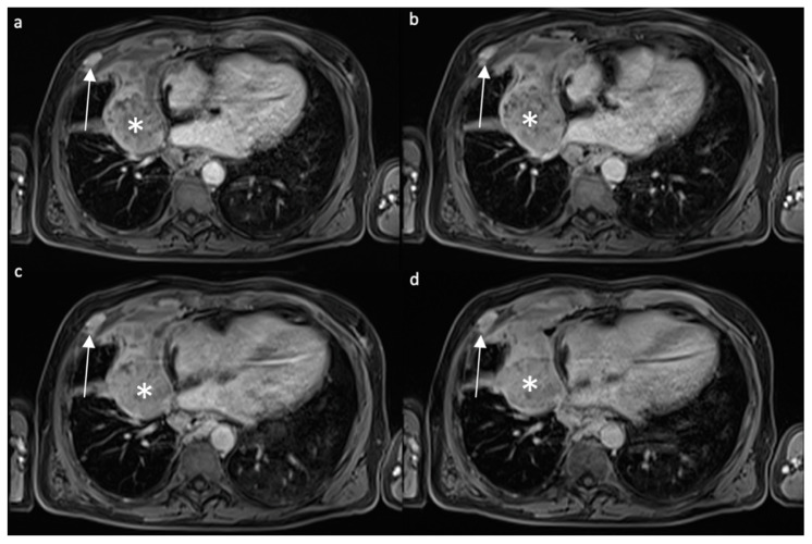
Figure 3.
Contrast-enhanced T1w axial fat-saturated images acquired with different timing after contrast administration ((a) 40 s, (b) 80 s, (c) 3′, (d) 5′) in a patient affected by MPM. White asterisk: paramediastinal mass with peripheral enhancement, in particular in the anterior portion; white arrow: enhancing nodule in the anterior thoracic wall is present.

