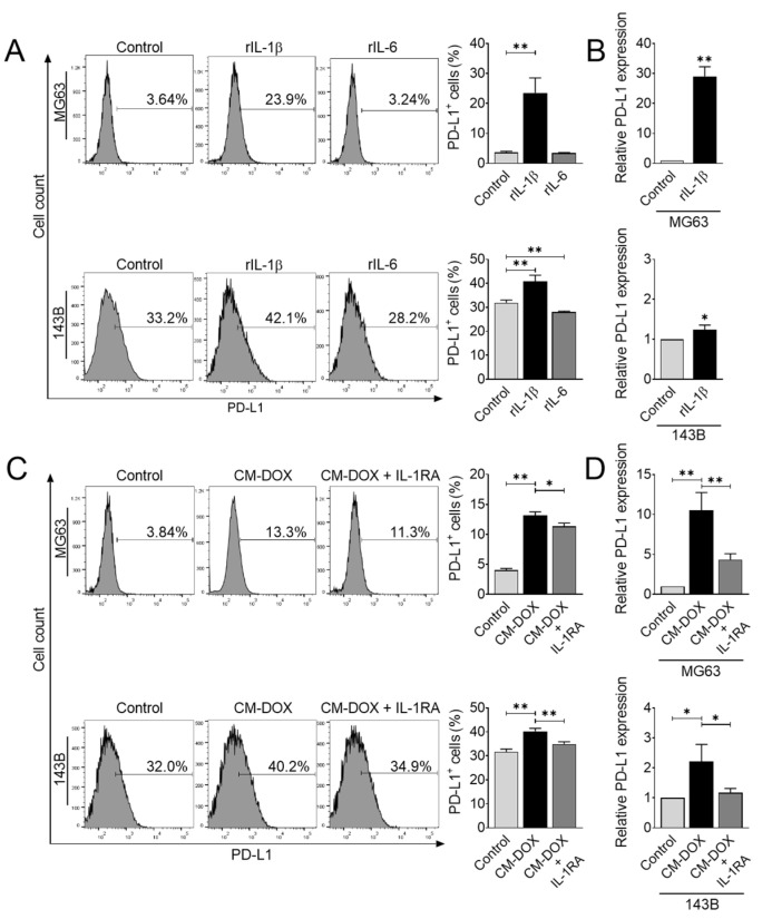
Figure 4.
IL-1β secretion in response to doxorubicin upregulates PD-L1 expression. (A) Flow cytometric analysis of PD-L1+ cells after recombinant IL-1β or IL-6 treatment. Human osteosarcoma cells were treated with rIL-1β (10 ng/mL) or rIL-6 (20 ng/mL) for 48 h, and PD-L1 protein expression was analyzed using flow cytometry. The data represent the means ± SD. **, p < 0.01 vs. control. (B) PD-L1 gene expression after rIL-1β treatment. Osteosarcoma cells were treated with rIL-1β (10 ng/mL) for 24 h, and qRT-PCR was performed to determine PD-L1 gene expression. The data represent the means ± SD. *, p < 0.05 and **, p < 0.01 vs. control. Flow cytometric (C) and qRT-PCR (D) analyses of PD-L1 expression after CM-DOX treatment. Osteosarcoma cells were treated with CM-DOX in the presence or absence of IL-1RA (100 ng/mL) and analyzed for PD-L1 expression. The data represent the means ± SD. *, p < 0.05 and **, p < 0.01 vs. control.

