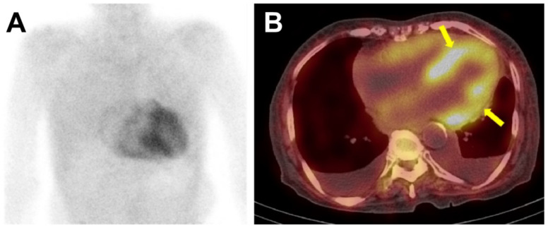
Figure 4.
The 99mTc-DPD bone scintigraphy and single-photon emission computed tomography (SPECT) of a patient with ATTR-cardiac amyloidosis. In Figure 4, the anterior planar image (panel A) showed intense, heterogeneous cardiac uptake greater than rib uptake in intensity, considered as Perugini grade 3. No abnormal radiotracer uptake was seen in other organs such as the liver or kidney. SPECT images (panel B) were acquired immediately after the planar imaging, and the radiotracer uptake could be localized to the myocardium (arrows), and not in the blood pool. Bilateral pleural effusion was also noted.

