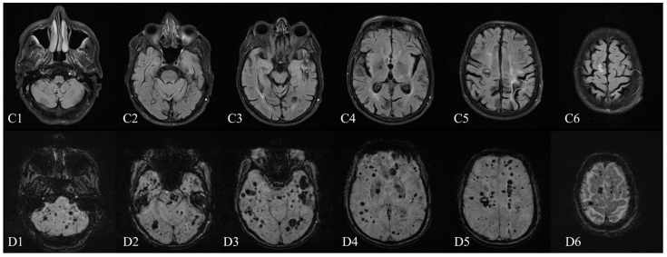
Figure 2.
Brain MRI (1.5 T)—January 2021: (C1–C6) axial FLAIR sequence and (D1–D6) axial SWI sequence (TE 40.00 ms). Compared with the 2015 MRI (1.5 T), the number and the dimensions of cavernomas substantially increased in almost every cerebral location (comparing the B1–B6 to the D1–D6 SWI series of imaging). The 2021 cerebral MRI (Figure 2) showed a significant increase in dimensions of many CCMs as well as the addition of novel lesions compared with the previous examination, without signs of acute bleeding. Some of the large CCMs (over 10 mm) identified on the 2015 MRI examination, of the Zabramski I and II subtype, had evolved to the Zabramski III subtype (Figure 3); moreover, countless new Zabramski type IV small lesions were observed [14]. The spinal examination revealed multiple focal lesions at cervical and thoracic levels, also highly suggestive of cavernous malformations (Figure 4C). One of the lesions, at the C3–C4 level, showed signs of recent hemorrhage (Figure 4A,B).

