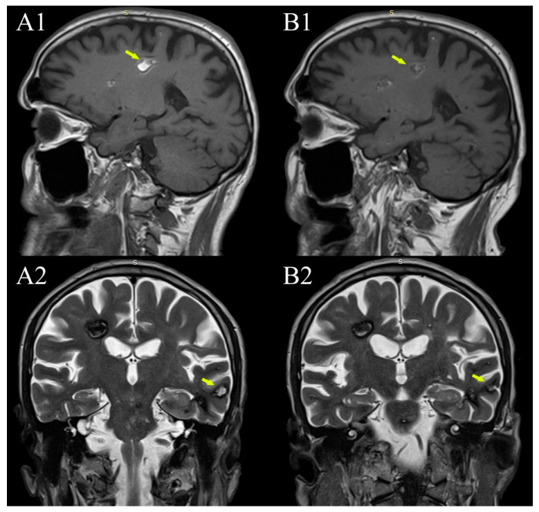
Figure 3.
Evolution of in-cavernomas bleeding over time. (A1,A2) are from the 2015 MRI examination (1.5 T), (B1,B2) are from the 2021 examination (1.5 T). Coexistence of subacute bleeding in the right parietal lobe white matter cavernoma with T1 hyperintense signal (A1—arrow) and T2 inhomogeneous hyper- and hypointense signal and acute bleeding in a smaller left temporal lobe cavernoma with hyperintense T2 signal (A2—arrow) (but also hyperintense T1, not shown here). Both lesions became more hypointense in both T1 and T2 on the 2021 MRI examination (B1,B2 —arrows).

