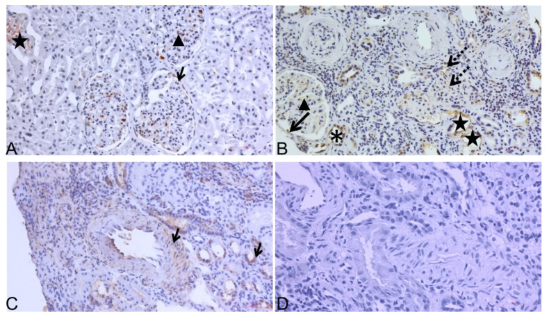
Figure 4.
ADAMTS-4 expression analyses by immunohistochemistry (IHC) in transplant kidney biopsy sample pair. (A) Representative staining of ADAMTS-4 in “zero-time“ sample (TX0) taken at time of transplantation: positive in GC, BC, and PXT; (B) representative staining of ADAMTS-4 in paired TXCI biopsy sample (stage CKD-4): positive in PTC and INT, where interstitial infiltrate is also seen along with GC and damaged PXT; (C) ADAMTS-4 positive control: positive in artery wall of middle sized intralobular artery (black arrow—left) and DT (black arrow—right); (D) secondary antibody only control in kidney biopsy sample was used. Original magnification 200×. DT, asterisk; BC, black arrow; GC, black arrow head; PTC, INT, dashed arrow; PXT, black star.

