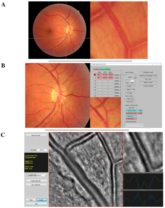
Figure 1.
Acquisition interface of retinal vessel measurements (A) using vessel analysis using VAMPIRE annotation tool, (B) IVAN software, or (C) adaptive optics camera and AO_Detect_Artery™. This figure briefly presents the acquisition interface of each vessel measurement technique that has been systematically used to measure four vessels (the two temporal arteries and the two temporal veins) of each retinal photograph. Vessel measurements were performed at the same section with all software tools. In this example the measured section of the superior temporal artery is framed in red.

