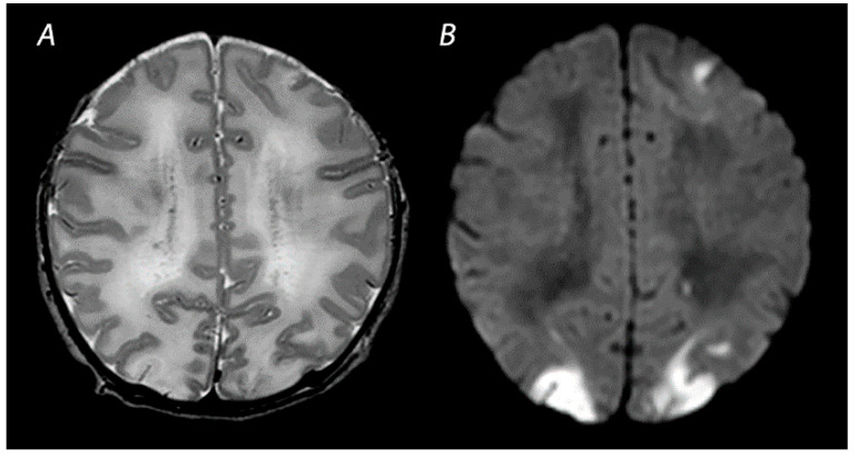
Figure 2.
MRI was performed on day 4 in a term infant who was born with Apgar scores of 1/5. Amplitude integrated EEG initially showed a continuous normal voltage background pattern and cooling was not started. The infant developed seizures at 10 h after birth. MRI shows a watershed predominant pattern of injury, with T2-weighted imaging showing loss of distinction between the gray and white matter at the cortex of the occipital lobes and the left frontal lobe (A) and DWI (B) showing diffusion restriction in these areas. The infant had a normal outcome at 18 months of age.

