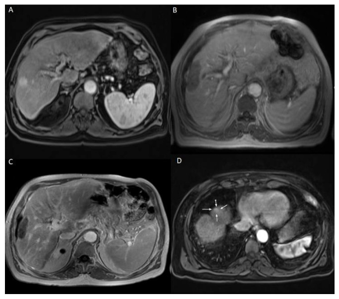
Figure 2.
69 years old male patient with chronic hepatitis C virus, liver cirrhosis, and HCC lesion in the right liver lobe, who was treated with LITT. (A) Pre-treatment CEMRI showed an arterial hyperenhancement of the lesion. (B) 24 h post-ablation CEMRI showed a large post-ablation zone, and the lesion was fully ablated. (C,D) At the 3-months post-ablation CEMRI, the size of the ablation zone was getting decreased without LTP, but there was a hyperenhancement in untreated liver segment (white arrows), which was correlated with IDR. TACE was performed to treat the recurrent HCC and the patient lived for 33 months after LITT until death.

