. 2022 Mar 7;11:e73360. doi: 10.7554/eLife.73360

Figure 3. TIGAR-deficient mice display increased ATP turnover in skeletal muscle.

(A) Wild type (WT) and whole-body Tigar knockout (TKO) male mice (n = 6) were maintained at room temperature (RT) or shifted to 4°C for 1 hr. Quadricep white skeletal muscles were isolated, and extracts assayed for ATP, ADP, and AMP levels as described in ‘Method details.’ (B) WT and TKO male mice (n = 3–4 per condition) were kept at RT or shifted to 4°C for 1 hr and then given an oral gavage of 0.3 ml pure H2O18 for 10 min at 4°C and an intraperitoneal (IP) injection of 1 ml pure H2O18 for another 10 min at 4°C. The quadricep muscles were freeze-clamped with liquid N2 and extracts prepared for mass spectroscopic analyses. The heatmap shows the O18 enrichment fraction of ATP, ADP, AMP, and creatine-phosphate. (C) WT and TKO male mice (n = 6) were maintained at RT or shifted to 4°C for 1 hr, and plasma creatine levels were determined by metabolomics analyses. (D) The CP_M4_83 data was generated from the same experimental setting as (B), indicating the increase in phosphocreatine turnover in TKO compared to that in WT at 4°C. Statistical analyses are described in ‘Method details,’ and the data are presented as the mean ± SD. *p<0.05, ***p<0.01.

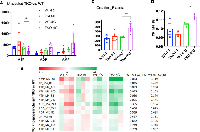
Figure 3—source data 1. Raw 18O enrichment data of stable isotope metabolic flux assessment with H218O was collected in quadricep white muscle of both ambient temperature housed and 4°C 1 hr exposed wild type (WT) and whole-body Tigar knockout (TKO) mice, as described in ‘Method details’.
The data was used in Figure 3.

Figure 3.

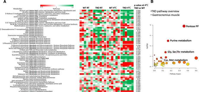
Figure 3—figure supplement 1. Skeletal muscles of whole-body Tigar knockout (TKO) mice at 4°C display increased pentose phosphate pathway, purine nucleotide cycle, and amino acid utilization pathways.

Figure 3—figure supplement 1.

Wild type (WT) and TKO male mice (n = 6) were maintained at room temperature (RT) or shifted to 4°C for 1 hr. (A) Quadricep white skeletal muscles were isolated, and extracts were subjected to widely targeted (multiple reaction monitoring [MRM]) small metabolite profiling using an ABSciex 6500 + QTRAP with ACE PFP and Merck ZIC-pHILIC columns as described in ‘Materials and methods.’ The heatmap shows the metabolites/pathways differentially identified with the corresponding p-values. (B) Matched pathways, determined from MetaboAnalyst 5.0, which integrates enrichment analysis (metabolite over-representation more than expected by chance) and pathway impact (reflecting the centrality of the metabolite in a topological network).
Figure 3—figure supplement 1—source data 1. Raw metabolites data were collected in the gastrocnemius muscle of both ambient temperature housed and 4°C 1 hr exposed wild type (WT) and whole-body Tigar knockout (TKO) mice as described in ‘Method details’.
The data was used in Figure 3—figure supplement 1.

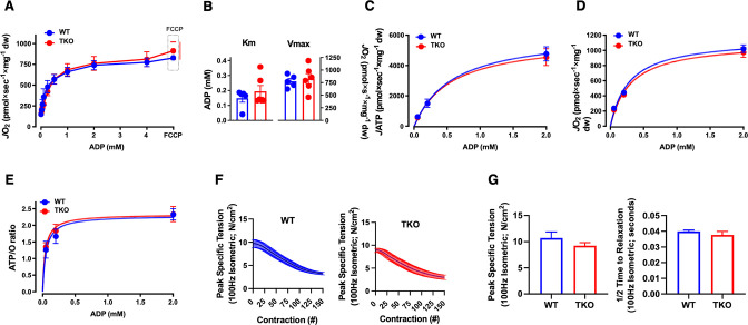
Figure 3—figure supplement 2. TIGAR deficiency has no significant effect on intrinsic skeletal muscle mitochondrial oxidative phosphorylation activity or ATP production.

Figure 3—figure supplement 2.

Permeabilized skeletal muscle fiber bundles were prepared from red portions of the gastrocnemius muscle from wild type (WT) and whole-body Tigar knockout (TKO) mice immediately after 1 hr cold exposure. (A) Oxygen consumption rate (JO2) as a function of ADP concentration. (B) The Km and Vmax for ADP-stimulatory respiratory kinetics. (C) Rates of ATP production (JATP) and (D) oxygen consumption (JO2) measured simultaneously under clamped submaximal and maximal ADP-demand states. (E) ATP/O efficiency ratios. (F) Force-frequency of the extensor digitorum longus muscle isolated from the WT and TKO mice. (G) Peak-specific tension and time to one-half relaxation of the extensor digitorum longus muscle from the WT and TKO mice.