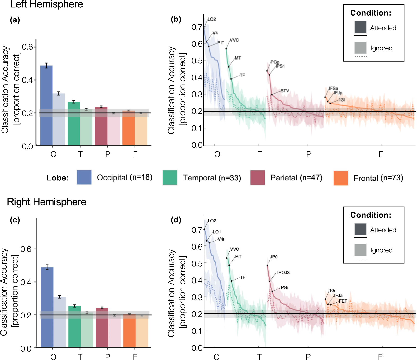
Fig. 3.

Classification accuracy by lobe, hemisphere, and condition. (a,c) Mean classification accuracy averaged across Glasser ROIs from each lobe: occipital (blue), temporal (green), parietal (red) and frontal (orange) lobes, in the left (a) hemisphere and right (c) hemisphere by condition: attended (dark colors) and ignored (light colors). Error bars: standard error of the mean across ROIs. (b,d) Same conventions as (a,c) but for each ROI separately. ROIs in the attended (solid line) and ignored (dashed line) conditions are ordered by the mean classification accuracy in the attended condition; shaded area: standard error of the mean across subjects. O: Occipital; T: Temporal; P: Parietal; F: Frontal. Horizontal lines : chance level; shaded region: the 95% confidence interval.
