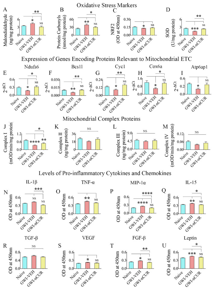
Figure 8.

Twelve weeks of nCUR treatment in rats with chronic GWI reduced oxidative stress, improved mitochondrial function, and repressed proinflammatory cytokines and chemokines in the hippocampus. The bar charts in A-D compare the concentration of malondialdehyde (MDA; A), protein carbonyls (PCs; B), the nuclear factor erythroid 2-related factor 2 (NRF2; C), and superoxide dismutase (SOD; D) in the hippocampus between naïve, and GWI rats receiving vehicle (GWI-VEH) or nCUR (GWI-nCUR). Note that MDA and PCs were upregulated, and NRF2 and SOD were downregulated in the GWI-VEH group, compared to the naïve control group. In the GWI-nCUR, all four markers were normalized to naïve control levels. The bar charts E-I compare the expression of genes Ndufs6 (E), Bcs1l (F), Cyc1 (G), Cox6a (H), and Atp6ap1 (I) between different groups. These genes' expression was downregulated in the GWI-VEH group but normalized to naïve control levels in the GWI-nCUR group. The bar charts in J-M compare the concentration of mitochondrial complexes I-IV in the hippocampus between different groups. The bar charts in N-U compare the concentration of interleukin-1 beta (IL-1β), tumor necrosis factor-alpha (TNF-α), macrophage inflammatory protein 1 alpha (MIP-1α or CCL3), interleukin-15 (IL-15), transforming growth factor-beta (TGF-β), vascular endothelial growth factor (VEGF), fibroblast growth factor-beta (FGF-β) and Leptin in the hippocampus between different groups. All of these cytokines and chemokines except IL-1β and TGF-β were upregulated in the GWI-VEH group but significantly reduced in the GWI-nCUR group. *, p<0.05, **, p<0.01, ***, p<0.001, and ****, p<0.0001; NS, not significant.
