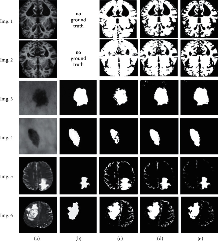
Figure 8.

Qualitative illustration for selected samples: (a) original images, (b) ground truths, (c) segmented images using the Gaussian-based MCET, (d) segmented images using the lognormal-based MCET, and (e) segmented images using the proposed MCET, the segmented MRI Alzheimer's Imgs 1.(e) and 2.(e) using alpha trim d/2 = 50, the segmented skin lesion Imgs 3.(e) and 4.(e) using alpha trim d/2 = 55, and the segmented MRI brain tumor Imgs 5.(e) and 6.(e) using alpha trim d/2 = 30.
