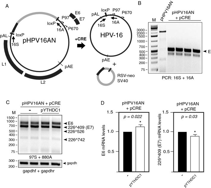
Fig. 5.
A Schematic representation of the pHPV16AN plasmid that carries the full-length HPV16 genome flanked by loxP sites. Co-transfection of pHPV16AN and pCRE results in excision and circulation of the viral DNA at the loxP sites to generate episomal HPV16 DNA. Positions of PCR primers 16A and 16S used to monitor HPV16 genome excision and circularization are indicated. B Gel shows PCR with primers 16A and 16S on DNA harvested 48 h post-transfection. E, episomal HPV16 DNA. C RT-PCR on RNA extracted from HeLa cells transfected with pHPV16AN and pCRE with empty pUC plasmid (−) or pYTHDC1. Transfections were performed in duplicates. RT-PCR was performed with the indicated RT-PCR primers. D Densitometric quantitation of gel images of RT-PCR products shown in (C). The RT-PCR band representing E6 mRNAs as well as the band representing spliced (226409) E7 mRNAs were quantified and fold difference in the absence or presence of YTHDC1 is shown in the graph. p-values are indicated

