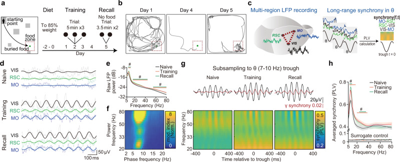
Fig. 1. Learning-induced cortical long-range gamma synchrony is coupled to the theta rhythm.
a A schematic of the spatial memory task. Mice were trained to find the place of hidden food (buried in sands) for 4 days. Recall trials were set on day 5 without any food reward. The arena was designed with landmarks (gray blocks) and buried food (green dot). Mice start exploring from the starting point in all trials. b Representative trajectories of one mouse during learning and recall. c Simultaneously recording of LFPs from three distinct cortices during the task (left) and calculation of phase-synchrony spectrogram by subsampling LFPs to theta trough (right). d Representative LFP raw traces (tint lines) and filtered traces (theta (θ) band, 7–10 Hz, dark lines) from three recording sites at different stages of the task. Here training shows the data from the first day of training, same as the following graphs. See results for each region for each day in the supplementary Fig. 1c. e, f Theta and gamma power increased in training/recall trials and the gamma power was coupled to cortical theta phase (78 electrodes from 28 mice, NMO = 26, NRSC = 26, NVIS = 26). e Averaged raw cortical LFP power from three regions of different trials. Encoding showed here is data from training day1. Significance was assessed by two-way ANOVA followed by false discovery rate (FDR) corrected multiple comparisons at each frequency comparing with data of homecage trail. The trial factor but not the frequency factor was repeatedly measured. Statistical significant frequency range (q < 0.05) is noted on the graph in corresponding colors. f Phase-power modulation index comodulograms of training, averaged from all regions. (See comodulograms of each region for each day and corresponding quantification in Supplementary Fig. 3). Modulation index was normalized by the element-wise division of the raw comodulogram by surrogated control. g, h Theta and gamma synchrony elevated in training/recall trials and the gamma synchrony was coupled to cortical theta phase (72 electrode pairs from 28 mice. NMO-RSC = 24, NRSC-VIS = 24, NMO-VIS = 24). g Gamma synchrony was specifically coupled to theta phase during training and memory recall. Top, averaged theta wave (black) and 30 Hz cortical synchrony (red). Bottom, averaged phase(theta)-synchrony(gamma) spectrogram from all pairs. Each pixel of the spectrograms showed averaged synchrony of all pairs. h Comparison of averaged overall synchrony in three kinds of trials (See result for each pair for each day in the supplementary Fig. 1d. q < 0.05, FDR corrected, significantly changed frequencies were noted on the graph).# q < 0.05, Shadow of line plot shows S.E.M.

