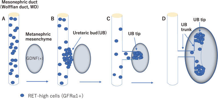
Figure 3.
RET-dependent cell movement during ureteric bud (UB) formation and branching. RET-positive cells (blue) are initially dispersed along the mesonephric duct (Wolffian duct, WD) (A). RET-positive cells start to move to form the primary UB and the ventral mesonephric duct is depleted of RET-positive cells (B). As the UB grows out, RET-positive cells form the UB tips while RET-negative cells follow and form the UB trunk (C, D).

