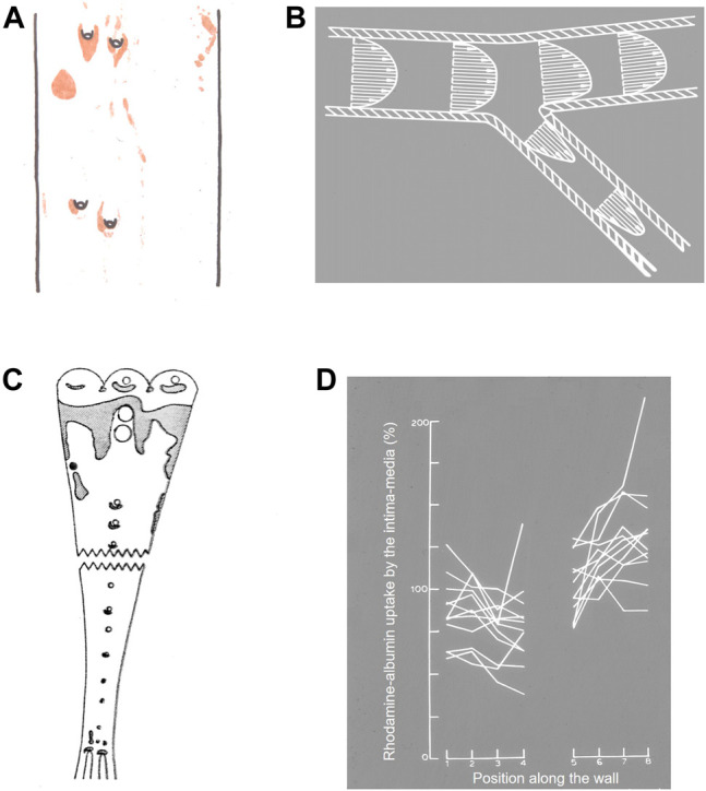
FIGURE 1.

(A) Watercolour from Anitschkow (1933) of lipid staining (red) near four intercostal branch ostia in a cholesterol-fed rabbit. The thoracic aorta is viewed en face and mean aortic flow is from top to bottom. (B) Conceptualisation of velocity vectors and velocity profiles in a 2-D model of a branching artery, after Fry (1969). (C) Uptake of Evans’ Blue Dye (shaded areas) in the aorta of a pig (Somer and Schwartz, 1971). Note that pigs have unpaired intercostal ostia, unlike the paired ostia in human and rabbit aortas. The aorta is viewed en face and mean aortic flow is from top to bottom. (D) Uptake of rhodamine-labelled albumin into the aortic wall of rabbits up to approximately one branch diameter upstream (positions 1→4) and downstream (positions 5→8) of 12 intercostal ostia (positions 4→5), 3 h after the tracer was administered (Weinberg, 1988).
