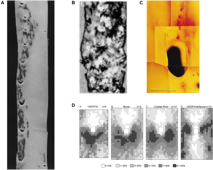
FIGURE 4.
(A) Human thoracic aorta, viewed as in Figure 2A, showing two patterns of lipid staining at intercostal branch ostia (Sloop et al., 1998). (B) Lesions in an aged human aorta, viewed as in Figure 2A. Fibrous caps of raised lesions appear white and can be seen completely surrounding the dark holes of branch ostia (Mitchell and Schwartz, 1965). (C) Spontaneous lipid deposition (red) extending upstream of the coeliac branch ostium in the abdominal aorta of an aged rabbit fed a normal diet (Barnes and Weinberg, 1998). (D) Maps of lesion prevalence, displayed as in Figure 3C, in mature rabbits of four different strains. The maps are arranged in an order that gives the impression of a trend from the downstream arrowhead to the lateral pattern of disease (Barnes and Weinberg, 2001).

