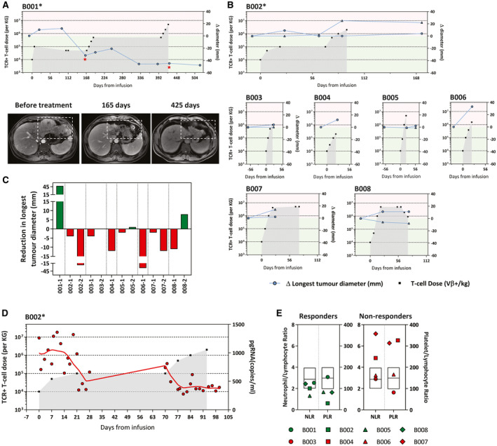
FIG. 4.

Immunological alterations in the peripheral blood correlate with observable antitumor response. The longest diameter of the target lesions in the liver was monitored throughout the treatment. The number of HBV‐specific TCR T cells infused are indicated as before. *Patients with reported adverse events. (A) Changes in the longest diameter (compared with baseline) of the liver target lesion in patient B001. Representative CT images of the liver target lesion at different time points are shown and indicated in the graph. (B) Changes in the longest diameter (compared with baseline) of the target lesion in the liver of patients B002‐B008. Each line represents a single liver target lesion. (C) Summary of the reduction in the longest diameter (compared with baseline) of individual liver target lesions in all treated patients analyzed from radiological imaging performed before treatment and within 3 months of the last infusion. (D) Levels of serum HBV pgRNA were analyzed in all patients. Only patient B002 (shown here) had detectable serum HBV pgRNA at baseline and was followed throughout treatment. (E) Baseline pre‐infusion neutrophil/lymphocyte and platelet/lymphocyte ratio of patients who exhibited immunological alterations after receiving HBV‐specific TCR T cells (responders) and those who do not (nonresponders). Box plot overlay shows the median and interquartile range of both parameters, as observed in the total population analyzed in Sangro et al.( 14 )
