
Figure 1.
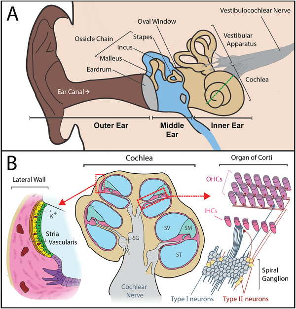
Anatomy of the ear and notable sites of degeneration that cause hearing loss. A) The outer ear collects and directs sound toward the tympanic membrane (eardrum). Soundwaves cause the eardrum to vibrate, and these vibrations are transferred to the bones of the ossicular chain (malleus, incus, and stapes) in the middle ear. This process amplifies the soundwaves. Amplified vibrations are then transferred to the inner ear, when the stapes footplate vibrates against the oval window of the cochlea. The inner ear consists of the cochlea and vestibular apparatus, where sound and movement are sensed respectively. The dotted green line represents the cochlear cross‐section displayed in (B). B) A cross‐section of the cochlea (center), which is a long spiraling tube, divided into three compartments. The scala vestibuli (SV) and scala tympani (ST) contain perilymph (138 mM Na+, 6.9 mM K+) and the scala media (SM) contains endolymph (2 mM Na+, 145 mM K+).[ 12 ] There are three main sites in the cochlea that can cause sensorineural hearing loss when damaged: 1) the stria vascularis (magnified left) which maintains the cochlear electrochemical gradient by recycling potassium ions (K+) and provides a protective blood‐labyrinth barrier, 2) the organ of Corti (magnified upper right), which contains the hair cells that convert sound into an electrochemical signal, and 3) the spiral ganglion (SG) neurons (magnified lower right), which convey auditory signals via the cochlear nerve to the brainstem. Type I spiral ganglion neurons receive auditory signals from the inner hair cells and convey these signals via the cochlear nerve to the brainstem, and type II neurons provide efferent signals from the brain. Type I neurons primarily innervate inner hair cells (IHCs) and type II neurons innervate outer hair cells (OHCs) (reviewed in ref. [13]).