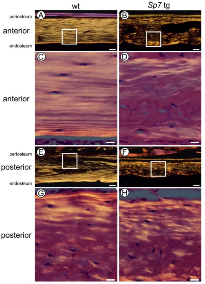
Figure 3.
Polarized microscopy of the cortical bone at diaphyses of femurs in male wild-type and Sp7 tg mice at 14 weeks of age: Anterior (A,B) and posterior (E,F) cortical bone in the wild-type (A,E) and Sp7 tg (B,F) mice. The boxed regions in (A,B,E,F) are magnified in (C,D,G,H), respectively. Scale bars = 50 μm (A,B,E,F); 10 μm (C,D,G,H). A total of seven wild-type and three Sp7 tg mice were analyzed and the representative pictures are shown.

