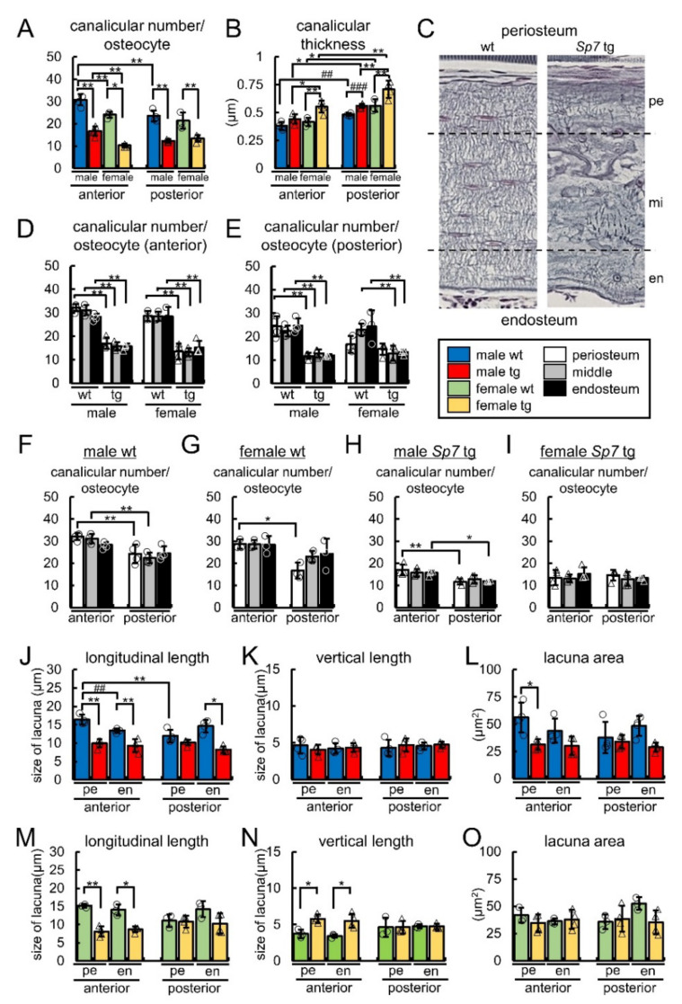
Figure 5.
The canalicular number, canalicular thickness, and the size of the lacunae in the diaphysis of the anterior and posterior femoral cortical bone in the wild-type and Sp7 tg mice of both sexes: (A) The average number of canaliculi in one osteocyte. (B) Canalicular thickness. (C–E) The average number of canaliculi in one osteocyte in three divided regions of the anterior (D) and posterior (E) cortical bone. The number of canaliculi from lacunae with osteocytes was counted in three divided regions, one-fourth area of the periosteum side (pe), half area in the middle (mi), and one-fourth area of the endosteum side (en), as shown in (C), and the average number of canaliculi in one osteocyte was calculated (D,E). A total of 12 groups were compared in (D,E). (F–I) Based on the data of (D,E), the canalicular number was compared between the anterior and posterior cortical bone in the three divided regions in the male wile-type (F), female wild-type (G), male Sp7 tg (H), and female Sp7 tg (I) mice. (J–O) The longitudinal length (J,M), vertical length (K,L), and area (L,O) of lacunae in the periosteal (pe) and endosteal (en) halves of the anterior and posterior cortical bone in the male (J–L) and female (M–O) wild-type and Sp7 tg mice. The number of mice that were analyzed; male wt and tg: 4, female wt: 3, female tg: 4. * p < 0.05, **,## p < 0.01, ### p < 0.001 by Tukey-Kramer test * and Student’s t-test #.

