Figure 3.
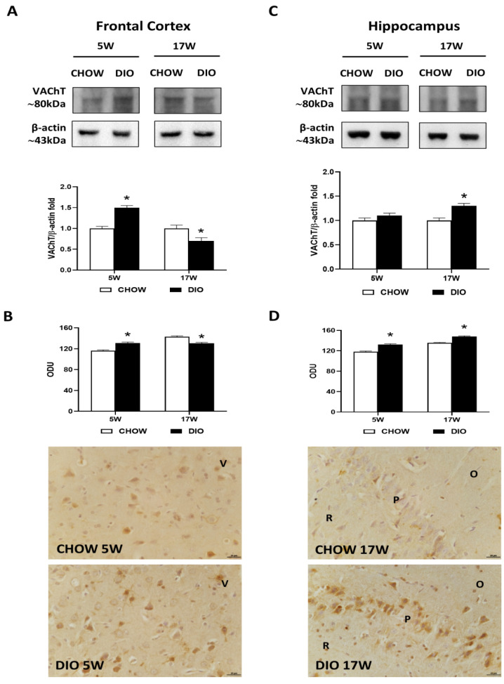
Western blot and immunohistochemistry of vesicular acetylcholine transporter (VAChT). Samples of the frontal cortex (A) and hippocampus (C) from rats fed with a standard diet (CHOW rats) and high-fat diet (DIO rats) for 5 and 17 weeks were immunoblotted with anti-VAChT. Graphs report the densitometric data with CHOW rats as control. ChAT membranes were stripped and incubated with anti-VAChT antibodies. β-actin control images were reused for illustrative purposes. VAChT immunoreaction intensity in the frontal cortex (B) and the hippocampus (D) from CHOW and DIO rats after 5 and 17 weeks of a diet was measured in optical density unit (ODU). Data are mean ± S.E.M. * p < 0.05 vs. age-matched CHOW rats. CHOW rats 5 weeks n = 8; DIO rats 5 weeks n = 8; CHOW rats 17 weeks n = 8; DIO rats 17 weeks n = 9. Representative pictures of CHOW and DIO rats frontal cortex after 5 weeks (B) and hippocampus, CA1 subfield, after 17 weeks (D) of diet. V, fifth layer of frontal cortex; O, stratum oriens; P, pyramidal neurons; R, stratum radiatum. Scale bar: 25 µm.

