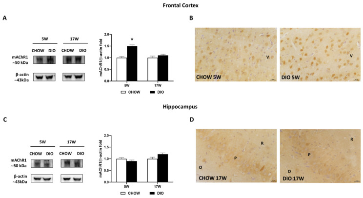
Figure 5.
Western blot and immunohistochemistry of muscarinic acetylcholine receptor subtype 1 (mAChR1). Samples of frontal cortex (A) and hippocampus (C) from rats fed with standard diet (CHOW rats) and high-fat diet (DIO rats) for 5 and 17 weeks were immunoblotted with anti- mAChR1. Graphs report the densitometric data with CHOW rats as control, and β-actin was used as a reference loading protein. Data are mean ± S.E.M. * p < 0.05 vs. age-matched CHOW rats. CHOW rats 5 weeks n = 8; DIO rats 5 weeks n = 8; CHOW rats 17 weeks n = 8; DIO rats 17 weeks n = 9. Representative pictures of CHOW and DIO rats frontal cortex after 5 weeks (B) and hippocampus, CA3 subfield, after 17 weeks (D) of diet. V, fifth layer of frontal cortex; O, stratum oriens; P, pyramidal neurons; R, stratum radiatum. Scale bar: 25 µm.

