Figure 3.
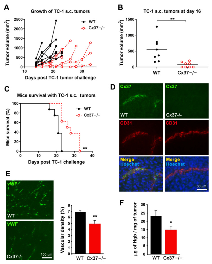
The growth of TC-1 tumors is reduced in Cx37−/− mice. (A) After s.c. injection, TC-1 cells generated growing tumors in all WT and Cx37−/− mice. (B) Sixteen days after the cell injection, the volume of tumors was significantly lower in Cx37−/− (n = 8) than in WT mice (n = 8). ** p < 0.01 versus WT mice (Student’s t-test). The horizontal bars show the mean tumor volume in each group. (C) All WT mice carrying the TC-1 tumor were sacrificed within the first 3 weeks of the experiments. In contrast, the cognate Cx37−/− mice survived significantly longer. ** p = 0.01 versus WT mice (log-rank Mantel-Cox test). (D) Sixteen days after the injection of TC-1 cells, Cx37 was detected via immunostaining in CD31-positive EC of the tumors grown in WT (left) but not Cx37−/− mice (right). Bar, 30 µm. (E) Immunostaining of the EC-specific von Willebrand factor (vWF) revealed a lower density of vessels in the tumors grown in Cx37−/− than in WT mice. Data are mean + SEM of 5–7 areas from size-matched tumors that developed in 4 mice per group. ** p < 0.01 versus WT mice (Student’s t-test). Bar, 100 µm. (F) The hemoglobin concentration was also lower in the tumors that developed in Cx37−/− than WT mice. Data are mean + SEM. n = 6 mice per group. * p < 0.05 vs. WT mice (Student’s t-test).

