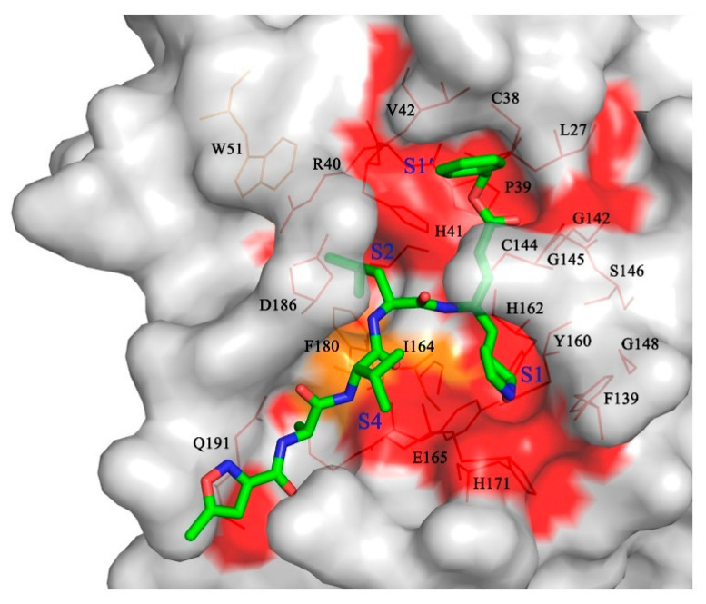
Figure 4.
Structure-based substrate-binding pocket conservation analysis of Mpros from four different genera. Surface representation showing the conserved substrate-binding pockets from the ten CoV Mpros listed in Figure 2B. The background is PDCoV Mpro. Red: identical residues among all ten CoV Mpros; orange: substituted in two CoV Mpros. The S1, S2, S4, and S1′ pockets and the residues that form the substrate-binding pocket are labeled. N3 is shown in green.

