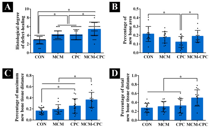
Figure 7.
Histomorphometric analysis of new bone formation. (A) histological evaluation of the degree of defect healing according to Huo et al. [19] based on hematoxylin and eosin and Masson–Goldner-trichome stained sections, (B) percentage of new bone formation area related to the initial defect area, (C) percentage of maximum distance of new bone to the initial defect length, (D) percentage of total distance of new bone to the initial defect length (mean ± SD, * p < 0.05).

