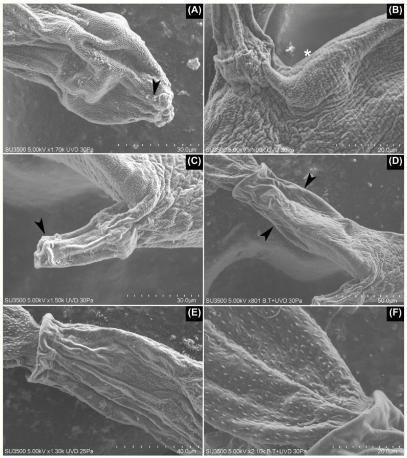
Figure 2.
SEM images of Lineage I. (A) The penetration organ with a pair of papilla-like structures on the border of the organ (arrowhead). (B) The body cuticle with a porous appearance (asterisk), clearly seen posterior to the acetabulum. (C) The acetabulum is covered on its tip with small, densely grouped spines (arrowhead), which contrasts with the rest of the acetabulum where no spines were seen. (D,E) Posterior third of the body where spines gradually disappear (arrowheads) until they are not seen on the posterior border. (E) Additionally, note the larger spines of the tail stem in comparison with those from the body. (F) The tail stem with a delicate honeycomb-shaped mesh over its tegument.

