Figure 3.
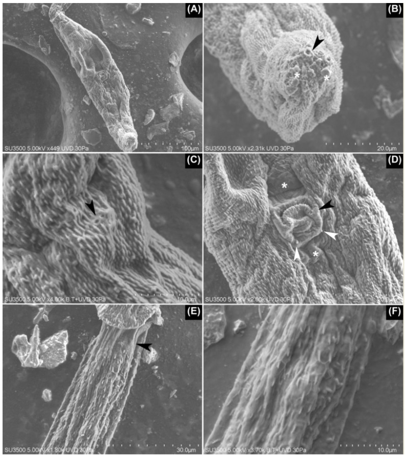
SEM images of Lineage II. (A) Ventral view of the body of furcocercariae completely covered with small spines over its tegument. (B) Apical view of the penetration organ with its glandular apertures (arrowhead) and some glandular content over these (asterisks). (C) The basal portion of the penetration organ where these two apertures were seen likely bore small cilia (arrowhead). (D) Inverted acetabulum with some small spines over its border (black arrowhead). Note the presence of two patches lacking spines—one anterior and the other posterior to the acetabulum (asterisks). At the basis of acetabulum, a pair of papillae-like structures are seen (white arrowheads). (E) At the anterior third of the tail stem, a small area lacking spines is evident (arrowhead). (F) Detail of the delicate, honeycomb-shaped mesh over the tegument of the tail stem, which is also seen in (E).

