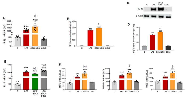
Figure 3.
CCL2 induces production of astrocytic inflammatory markers. (A) Astrocyte cultures were incubated with or without CCL2 (100 ng/mL). Twenty-four hours later, the whole medium was replaced in all wells, maintaining CCL2 where it was used for pre-treatment and adding LPS (0.1 g/mL) to indicated groups. Forty-eight hours later, RNA was isolated and IL-1β mRNA levels determined by RT-PCR. Data are means ± SEM of n = 6 replicates per group. *** p < 0.001 vs. C-C. ϕ p < 0.05 vs. LPS. (B) IL-1β concentration in the media was measured by ELISA. Data are means ± SEM of n = 6 replicates per group. *** p < 0.001 vs. C. ϕ p < 0.05 vs. LPS. (C) Cytosolic lysates were examined for the presence of IL-1β and β-actin protein by Western blot analysis. The gels shown are representative of experiments done on three separate astrocyte preparations. (D) Densitometric analysis of the bands. AU: arbitrary units relative to C. *** p < 0.001 vs. C. ϕ p < 0.05 vs. LPS. (E) Astrocyte cultures were incubated with vehicle (EtOH 0.037%), RvD1 100 nM (dissolved in EtOH 0.037%) or RvD1+CCL2 (100 ng/mL). Twenty-four hours later, the whole medium was replaced in all wells, maintaining RvD1 and CCL2 where it was used for pre-treatment and adding LPS (0.1 g/mL) to all groups except the control one. Forty-eight hours later, RNA was isolated and IL-1β mRNA levels determined by RT-PCR. Data are means ± SEM of n = 6 replicates per group. *** p < 0.001 vs. C. ϕϕ p < 0.01 vs. LPS. δδδ p < 0.001 vs. LPS+RvD1. (F) Astrocyte cultures were incubated with or without CCL2 (100 ng/mL). Twenty-four hours later, the whole medium was replaced in all wells, maintaining CCL2 where it was used for pre-treatment and adding LPS (0.1 g/mL) to indicated groups. Forty-eight hours later, RNA was isolated and TNFα, MIP1α and NOS2 mRNA levels determined by RT-PCR. Data are means ± SEM of n = 6 replicates per group. *** p < 0.001 vs. C-C. ϕϕϕ p < 0.001, ϕ p < 0.05 vs. LPS.

