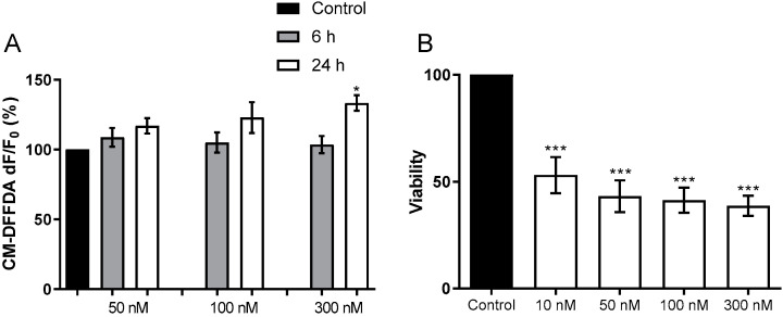
Figure 2.
Toxicity of HPU to BV-2 cells (A) For ROS detection, BV-2 cells were incubated CM-DFFDA (2 mM) probe for 30 min, 37 °C, in the dark. After washing, BV-2 cells were incubated with 20 mM NaPB (control), or 50, 100 or 300 nM HPU for 6 h (gray columns) or 24 h (white columns). Fluorescence was measured at λex495 nm/λem527 nm. The results were expressed as a percentage of the control ± SEM and compared by one-way ANOVA followed by the Tukey test. * p < 0.05 vs. controls. (B) BV-2 viability was analyzed by the MTT test after 24 h of exposure to HPU. The cultures’ supernatants were removed after the treatments and cells were incubated with MTT (5 mg/mL) for 4 h at 37 °C, then suspended in 100 µL DMSO. Absorbances were read at 570 nm. Mean ± SEM. *** p < 0.001 vs. control.

