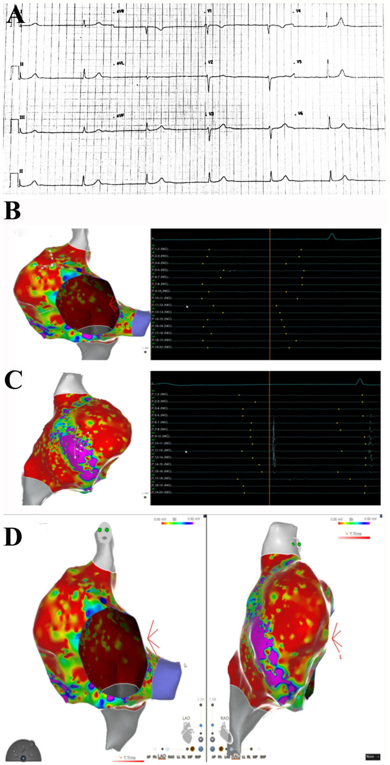
Figure 1.
Right atrial substrate mapping and endocavitary electrogram recordings. ECG shows no sinus rhythm, no atrial activity at EPS. Junctional rhythm at 40 bts/min (A). Endocavitary recordings show the absence of atrial activation during atrial mapping (B). Some atrial signals are recorded on the anterolateral wall of right atrium (C). Left and right anterior oblique projections of right atrium endocardial substrate mapping shows a large scarred area with low-voltage signals (<0.05 mV, in red), with a small portion of myocardial wall with preserved voltage signals (>0.5 mV, in purple) on the anterolateral aspect of right atrium (D).

