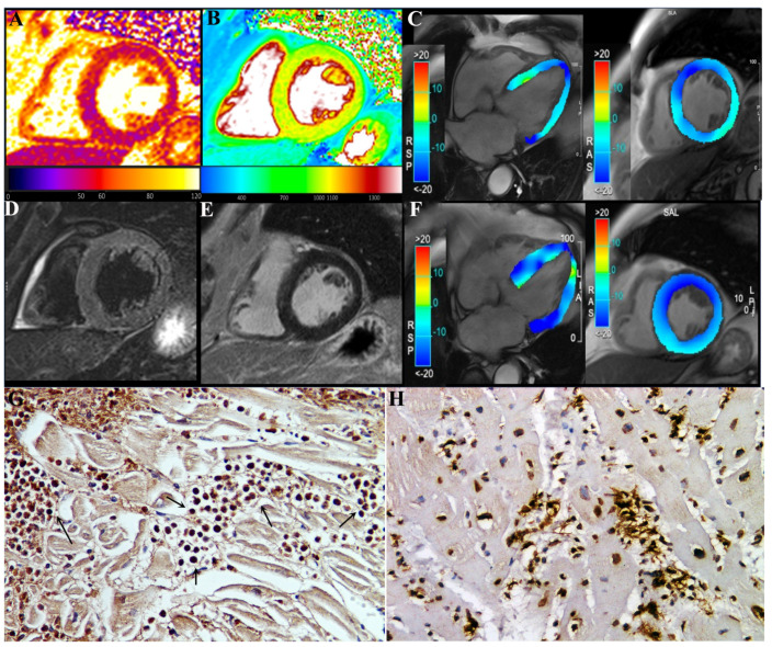
Figure 3.
A 78-year-old male (pt2) with eosinophilic myocarditis myocarditis after second dose of COVID-19 mRNA (BNT162b2) vaccine. At first CMR exam (A–E), performed 4 weeks after symptoms onset, T2 (A) and native T1 (B) maps revealed an increase in global T2 (55 ms ± 4 ms, normal range < 49.9 ms) and T1 (1060 ± 15 ms, normal range < 1027 ms) myocardial values, without any noticeable focal areas of signal changes on STIR T2-weighted (D) and late-gadolinium-enhanced (E) images, reflecting mild diffuse edematous condition. CineMR images, acquired on four-chamber (left) and mid-ventricular short-axis (right) views, analyzed by tissue tracking analysis show moderate impairment of both longitudinal and circumferential systolic left ventricular function (GLS: −12.3%, GCS: −13.6%, EF: 35%) at admission (C), which improved at two-month follow-up after therapy ((F), GLS: −18.1%, GCS: −17.8%, EF: 49.5%). GCS: global circumferential strain; GLS: global longitudinal strain; EF: ejection fraction. (G) shows in post-vax myocarditis massive eosinophilic myocardial infiltration with necrosis of the adjacent myocytes (IHC for eosinophil major basic protein (EMBP) antibody). (200× magnification). (H): Comparison with acute myocarditis associated to COVID19 infection showing intense lymphocytic inflammation (IHC for CD45Ro) (200× magnification).

