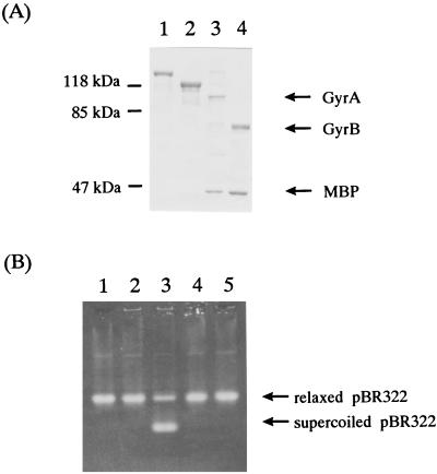
FIG. 4.
Purification of B. fragilis GyrA and GyrB proteins. (A) Sodium dodecyl sulfate-polyacrylamide gel electrophoresis analysis of purified B. fragilis GyrA and GyrB proteins. The proteins were electrophoresed in a 10% polyacrylamide gel and stained with Coomassie brilliant blue. The masses of the protein markers are indicated in kilodaltons on the left. Lane 1, MBP-GyrA fusion protein; lane 2, MBP-GyrB fusion protein; lane 3, MBP-GyrA fusion protein after factor Xa cleavage; lane 4, MBP-GyrB fusion protein after factor Xa cleavage. (B) Supercoiling activity of purified GyrA and GyrB proteins. Lane 1, purified GyrA (1 U); lane 2, purified GyrB (1 U); lane 3, purified GyrA (1 U) and GyrB (1 U); lane 4, purified GyrA (1 U) and GyrB (1 U) without ATP; lane 5, no addition. The source of DNA is pBR322.

