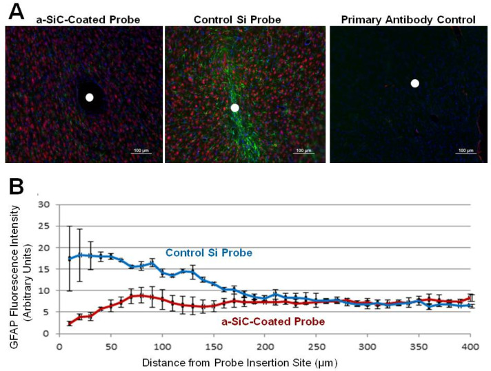
Figure 5.
(A) Images from deep cortical tissue labeling NeuN in red (neurons), GFAP in green (astrocytes), and DAPI in blue (cell nuclei). White circles indicate center of probe locations. Left image is from tissue implanted with an a-SiC-coated probe, middle from a control Si probe, and the right image is the no primary control, indicating little or no non-specific antibody binding. (B) Immunohistochemistry summary data comparing results from a-SiC-coated (red) and control Si (blue) probe after implantation for four weeks. Data are mean ± SEM for n = 2 slices [3].

