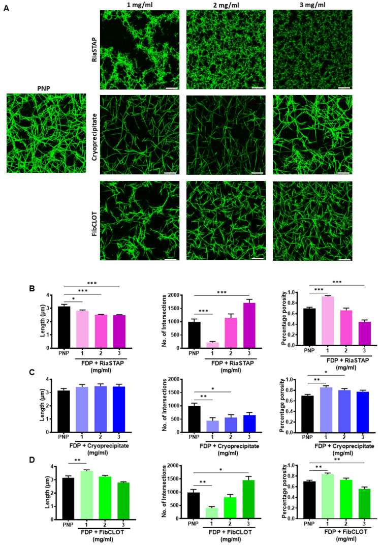
Figure 3.
Impact of fibrinogen source on fibrin network structure. Clots were formed containing 30% PNP or FDP, 0.25 µM AF488 fibrinogen, 16 µM phospholipids ± RiaSTAP®, cryoprecipitate or FibCLOT. Clots were polymerized by addition of 0.1 U/mL thrombin and 10.6 mM CaCl2 for 2 h at 37 °C. Z-stack images were collected on an LSM 710 confocal microscope using a 63 X oil objective. Scale bar = 20 µM. (A) Images representative of n = 3. Data shown are the mean ± SEM fibrin fiber length (μm), number of intersections present indicating branching and the percentage porosity of the clots for (B) RiaSTAP® (C) cryoprecipitate and (D) FibCLOT®. * p < 0.05, **p < 0.01 and *** p < 0.001.

