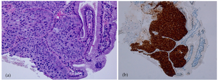
Figure 2.
Histopathology image of CIN3/HSIL. The epithelium lacked maturation and consisted of highly atypical cells with hyperchromatic nuclei with increased mitotic activity. The nuclear/cytoplasmic ratio was also increased. (a) Hematoxylin and eosin staining (magnification 100×). (b) Immunohistochemical staining with p16 revealed a diffuse reaction, which was highly suggestive of CIN3 (magnification 100×).

