Figure 5.
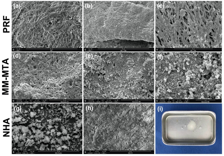
Representative scanning electron microscope images at 3000× magnification (a,b,d,e,g,h) and 12,000× magnification (c,f). The morphology observed for (a) PRF and (d) for MM-MTA and (h) NHA before immersion in PBS; (g) NHA powder; (b,c) the morphology observed for PRF, (e,f) for MM-MTA and (i) for NHA after immersion in PBS for 7 days at 37 °C.

