Figure 1.
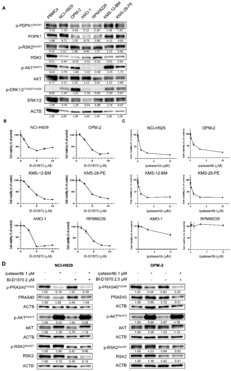
Activities and the role of cell proliferation of RSK2 and AKT in human myeloma–derived cell lines (HMCLs): (A) Baseline activities of RSK, AKT, and related kinases examined by Western blot (WB) in six HMCLs and peripheral blood mononuclear cells (PBMCs). The expression level of ACTB was examined as the internal control. Expression levels relative to ACTB are shown below each band measured by densitometric analysis using Image-J software. (B,C) Growth inhibitory effects of BI-D1870 (C) or ipatasertib (D) in six HMCLs. Cells were seeded at 2 × 10⁵ cells/mL and treated with various concentrations of BI-D1870 (C) or ipatasertib (D) for 48 h. The IC50 values of BI-D1870 for NCI-H929, OPM-2, KMS12-BM, KMS28-PE, AMO-1, and RPMI8226 cells were 4.00, 4.34, 3.88, 4.95, 3.14, and 4.09 μM, respectively, while those of ipatasertib for NCI-H929, OPM-2, KMS12-BM, and KMS28-PE cells were 0.95, 2.12, 0.81, and 0.79 μM respectively. (D) Effects of BI-D1870 and ipatasertib on their target molecules in NCI-H929 and OPM-2 cells. Cells were treated with either ipatasertib, BI-D1870, or their combination at the indicated concentrations for 48 h. Expression levels relative to control (untreated cells) are shown below each band measured by densitometric analysis using Image-J software. ACTB was used as an internal control.

