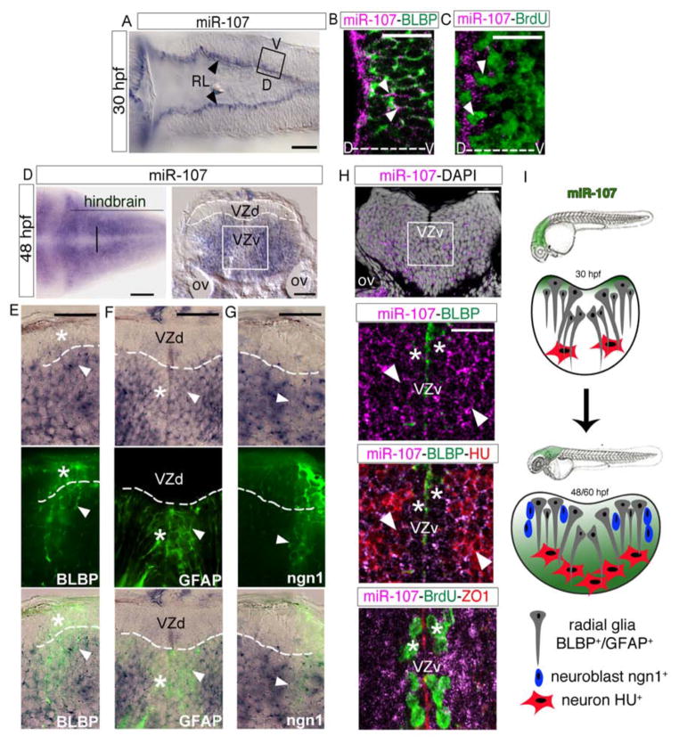
Figure 1. miR-107 is expressed in newborn neurons and in neural/neuronal progenitors across the hindbrain periventricular zone.
(A) Expression of miR-107 at 30hpf is restricted to the rhombic lip (RL) (arrowheads). Dorsal is up and anterior to the left. (B–C) Whole-mount confocal images of the boxed area in (A), stained with miR-107 LNA probe and anti-BLBP antibody in (B) or anti-BrdU antibody in (C). Brain orientation is indicated as D (dorsal) and V (ventral). Arrowheads indicate miR-107 expression. (D–H) Expression of miR-107 across the hindbrain of 48 hpf embryos. (D) Bright-field images of whole mount embryos (left panel) and hindbrain transverse section (right panel). VZd indicates the dorsal ventricular hindbrain zone (white dotted line), VZv indicates ventral ventricular hindbrain zone (white solid square). (E–G) High magnification images (60X) of the VZd zone representative of the region highlighted in D (left panel). miR-107 and protein co-localization is indicated with arrowheads, while miR-107 negative or low expressing cells are indicated with stars. (H) Top panel is a confocal image of a transverse hindbrain section at the level of the otic vesicle (OV). miR-107 is shown in purple and DAPI nuclei are in white. Stars indicate miR-107 expression in the ventral ventricular zone (VZv). Bottom panels are coronal confocal sections at the VZv level highlighted by the white box in the top panels and stained for the indicated proteins. ZO1 staining indicates the apical region of the VZv where BrdU positive cells are nested (white stars). (I) Schematic representation of the dynamic expression of miR-107 (green) in the developing hindbrain from 30 to 48/60 hpf. Scale bars= 50μm.

