
Extended Data Fig. 4. Comparison of the Cbl-binding, RS, and C-terminal domains of TokK, KsTsrM, and OxsB.
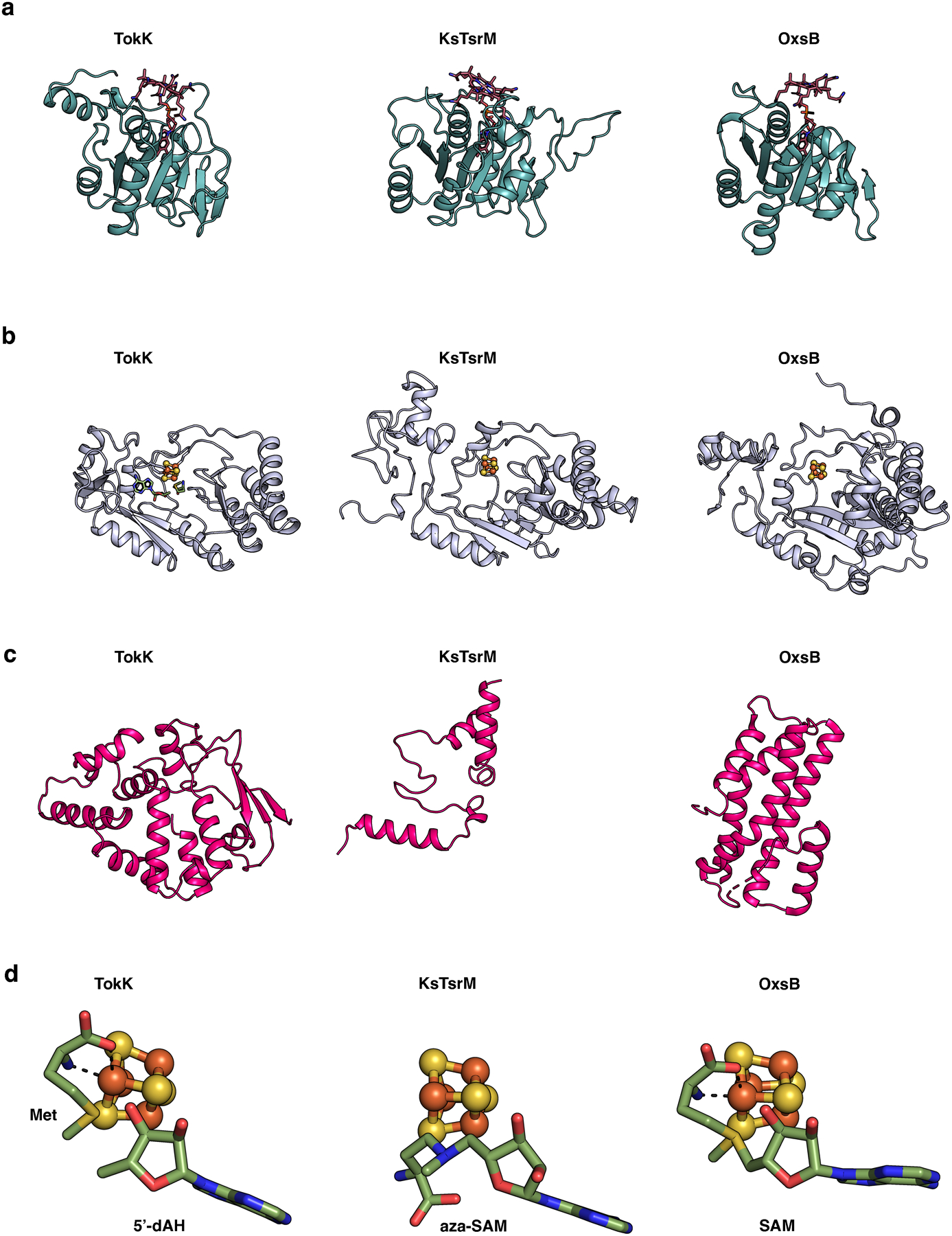
The domains are colored as in Extended Data Fig. 3. (a) The Rossmann fold, in teal, is highly similar among TokK (PDB ID: 7KDY), KsTsrM (PDB ID: 6WTF), and OxsB (PDB ID: 5UL4). (b) The core of each of the RS domains is a (β/⍺)6 motif; however, there are distinct differences. The RS domain of OxsB is more compact than those of TokK or KsTsrM, and all three have unique extra secondary structure features. (c) The C-terminal domains for TokK, KsTsrM, and OxsB are vastly different in architecture. (d) Comparison of the binding of Met and 5’-dAH, aza-SAM, and SAM for TokK, KsTsrM, and OxsB structures, respectively. Only the relevant binding of SAM to the cluster is shown for OxsB. OxsB has two binding positions of SAM, one to the cluster and one in what is proposed to be an intermediate state towards methylating the Cbl.