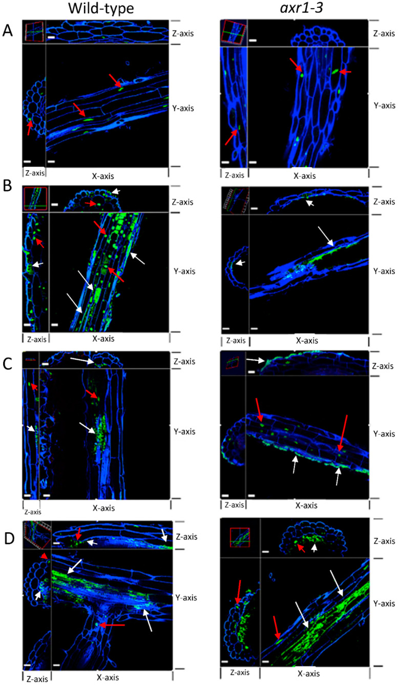
Figure 5.
IAA-producing strains that inhibit root length differentially colonize axr1-3 root tissue. Confocal microscopy showing orthogonal view of wild-type (left panels) and axr1-3 (right panels) roots upon treatment with (A) no bacteria, (B) IAA-producing Microbacterium RU1A, (C) IAA-producing Azospirillum baldaniorum Sp245, and (D) low IAA-producing Herbaspirillum RU5E that does not inhibit root length. The microscopy channels for each dye are blue (calcofluor white used to stain cell wall) and green (SYBR Gold DNA used to stain the nucleus and bacteria). White arrows indicate bacteria locations based on the size and morphology of the DNA-stained spots. Bacteria are shown as green spots that are smaller in size to plant nuclei (shown with red arrows) and tend to form clusters. The size bar in white represents 20 μm on each panel. The 3D images are rotated at the z-axis at two different locations of the tissue shown (top and left sections of each panel as shown by the cross-hair in the upper left corner image) to illustrate transverse views at the location of the stained spots and demonstrate either epiphytic or endophytic locations.

