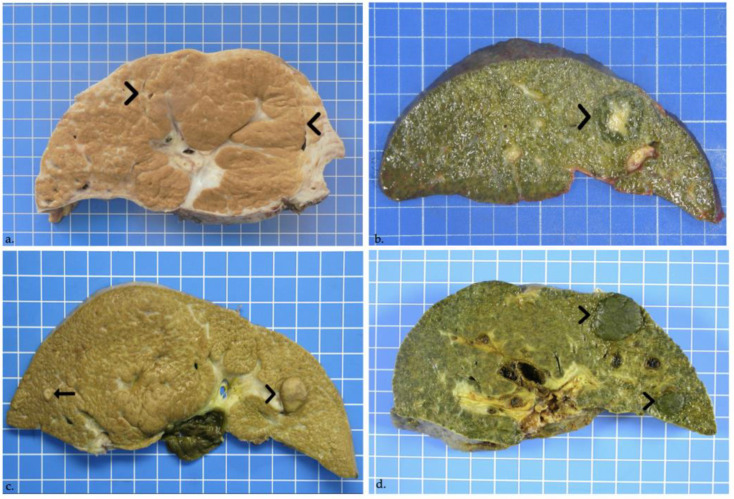
Figure 3.
(a) Regenerative nodule in a male patient 7-years-old at liver transplantation. On cut section, a rather ill-defined 8-cm large regenerative macronodule involves segments IV, V and VIII (delineated by arrowheads). (b) Focal nodular hyperplasia in a 13-month-old girl. On cut section, a 2.5-cm lobulated and well-defined though unencapsulated cholestatic lesion with a central scar is seen in segment IV (arrowhead). (c) High-grade dysplastic nodule in a female patient aged 3 years and 5 months at transplantation. Macroscopy shows a 1.5-cm bulging brown nodule in liver segment III (arrowhead), and a further 0.4-cm nodule in segment VI (arrow). (d) Well-differentiated hepatocellular carcinoma. Two large cholestatic nodules in segments II and III, measuring 2.3 and 1.1 cm in greatest diameters, bulge out from the cut section (arrowheads).

