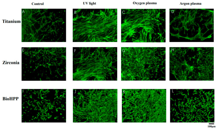
Figure 2.
Cell attachment and morphology of osteoinductive DPSCs on different surface treated titanium, zirconia and BioHPP after 21 days. The attachment of osteoinductive cells at 21 days of culture was observed using fluorescence microscopy (20×). Adhesion of cells was weak and cells were loosely distributed on untreated surfaces of titanium, zirconia and BioHPP (A,E,I). Either UV light or NTP treatment resulted in stronger cell adhesion and dense distribution (B–D,F–H,J–L), whereas argon plasma treatment led only to sparse cell adhesion (D,H,L).

