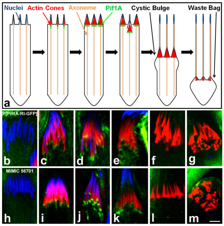
Figure 10.
Pif1A localization time series. Schematic of localization of Pif1A protein throughout spermatid individualization (a); Pif1A (green) is present in the front region of nascent needle-shaped actin cones and is not observed in individualized cones. Representative homozygote P[Pif1A-RI-GFP] individualization complexes (b–g) stained with DAPI (blue), phalloidin/actin (red) and Pif1A-GFP (green) and representative MiMIC-GFP cones (h–m) stained the same way—localization profile is consistent with homozygote Pif1A-GFP transgenic cones. Bar 5 µm.

