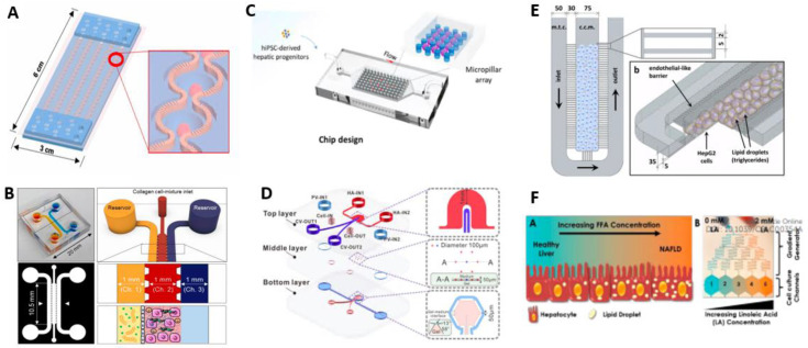
Figure 2.
Modeling NAFLD using microfluidic devices. (A) Steatochip recapitulating liver microstructure, for growth and differentiation of HepRG cells [54] (reproduced with permission from Teng et al., 2021). (B) NASH-on-chip, prepared with hydrogel channel with hepatic, Kupffer and stellate cells [55] (reproduced with permission from Freag et al., 2020). (C) NAFLD-on-chip inducing differentiation of hiPSC to organoids followed by steatosis progression [56] (reproduced with permission from Wang et al., 2020 Copyright: ACS Biomater. Sci. Eng. 2020, 6, 5734−5743). (D) Schematic illustration of liver lobule chip [57] (reproduced with permission from Du et al., 2021). (E) NAFLD on-on-a-chip in 3D and schematic view with HepG2 cell culture [58] (reproduced with permission from Gori et al., 2016; this is an open access article distributed under the terms of the Creative Commons Attribution License). (F) NAFLD model on chip generating the gradient of liloleic acid [59] (reproduced with permission from Bulutoglu et al., 2019).

