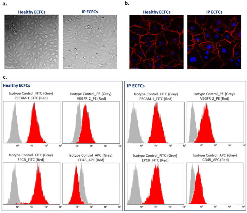
Figure 2.
Phenotype characterization of endothelial colony-forming cells (ECFCs). (a) Typical endothelial cobblestone-like morphology of ECFCs isolated from a healthy individual and the index patient (IP) observed using bright-field microscopy. Scale bars, 100 µm. (b) Expression of VE-cadherin (red) at cell–cell junctions is visualized with immunostaining of ECFCs derived from a healthy individual (left side) and patient (IP ECFCs) (right side). The nucleus is stained with DAPI (blue). Scale bars, 50 µm. (c) Flow cytometry analysis of ECFCs derived from a healthy donor (left images) and the IP (right images). The data confirmed that both healthy and IP isolated cells were positive for endothelial cell canonical markers of PECAM-1 (CD31-FITC conjugated), VEGFR-2 (PE-conjugate), and EPCR (FITC-conjugate); besides, they were negative for leukocyte cell marker CD45 (APC-conjugate). Isotype controls were conjugated with either FITC, PE, or APC corresponding to their relevant antibodies, and they are shown as grey bell curve graphs.

