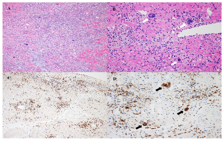
Figure 2.
Pathologic findings of the heart. Diffuse cardiomyocyte necrosis and mixed inflammatory infiltration are noted (A; H&E, ×100). Mixed inflammation including lymphocytes, macrophages, and frequent eosinophils are noted, and multinucleated giant cells are also noted (B; H&E, ×200). T-lymphocytic infiltration is identified (C; CD3, ×100). Infiltration of CD68+ macrophages is identified, and multinucleated giant cells are positive for CD68 (D; CD68, ×200). Black and white arrows show multinucleated giant cells.

