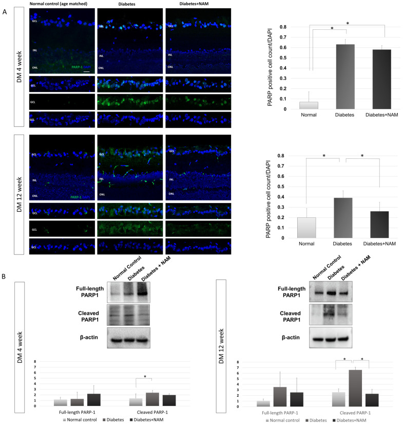
Figure 7.
(A) Poly(ADP-ribose)polymerase (PARP)-1 immunofluorescence staining. The proportion of PARP-1-positive cells was elevated in the diabetes group compared to the normal control group at 4 weeks and 12 weeks after onset of diabetes (both p < 0.001). An increased expression of PARP-1 in diabetic conditions was decreased by nicotinamide treatment at 12 weeks after induction of diabetes (p < 0.001). (B) Western blotting of poly(ADP-ribose)polymerase (PARP)-1. There was no significant difference in the expression of full-length PARP-1 according to the presence of diabetes or nicotinamide treatment at 4 or 12 weeks after onset of diabetes (p = 0.228, p = 0.103, respectively) Cleaved PARP-1 expression was elevated in the diabetes group compared to the normal control group at 4 and 12 weeks after induction of diabetes (p = 0.002, p < 0.001, respectively). Elevated expression of cleaved PARP-1 was reduced by nicotinamide treatment 12 weeks after onset of diabetes (p < 0.001). Scale bar = 20 μm. DM, diabetes mellitus; NAM, nicotinamide. GCL, ganglion cell layer. INL, inner nuclear layer. ONL, outer nuclear layer. *, significantly different as indicated (p < 0.05).

