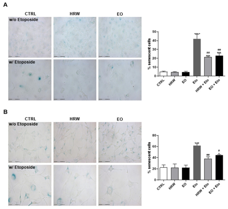
Figure 6.
Effect of HRW and EO extracts from E. globulus leaves on the senescence-associated β-galactosidase activity in etoposide (Eto)-stimulated HaCaT keratinocytes (A) and NIH/3T3 fibroblasts (B). Cellular senescence was induced using 100 µM Eto for 72 h for HaCaT and 12.5 µM Eto for 24 h for NIH/3T3. After the incubation period, the senescent cells were treated in the absence or presence of 0.16 mg/mL EO or 0.8 μg/mL HRW for 24 h. Cells treated with the medium alone were used as the CTRL. The senescent cells were quantified using a Senescence β-Galactosidase staining kit. The results—expressed as the percentage (%) of the senescent cells—represent the mean ± SEM of at least three independent experiments performed in duplicate. The statistical analysis was performed using one-way ANOVA, followed by Dunnett’s and Sidak’s multiple comparison tests. * p < 0.05, *** p < 0.001, and **** p < 0.0001: significantly different compared to the CTRL. # p < 0.05, and ## p < 0.01: significantly different compared to the Eto.

